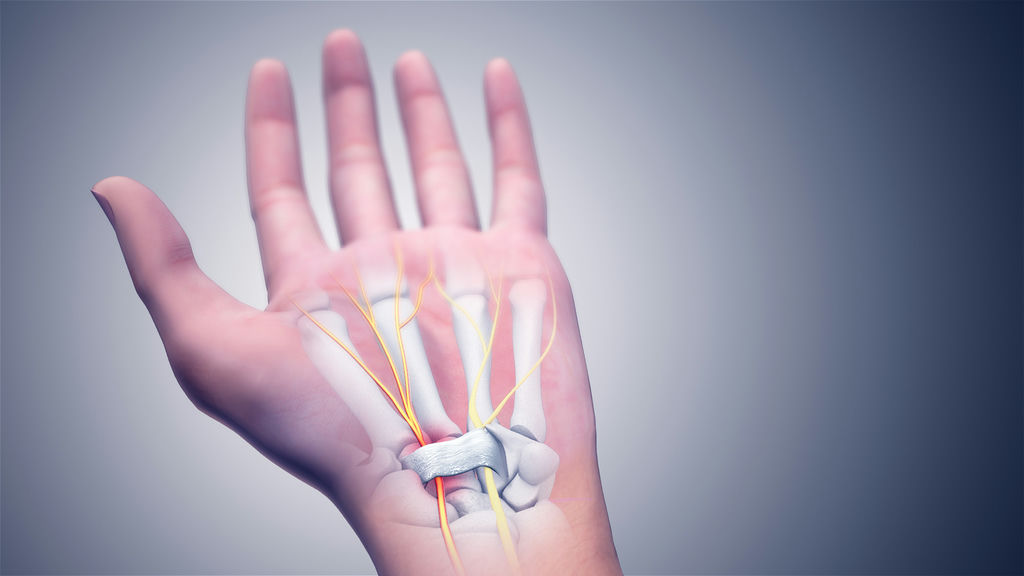
Carpal Tunnel Syndrome (CTS) affects millions of people, especially those who perform repetitive hand movements.
When it comes to surgical treatment, there are 3 leading techniques which are available:
All three techniques differ in more ways than one. Read more below to see the differences.
Dr. Thomaz Silva – About ultrasound-guided carpal tunnel release.
Ultrasound Guided Micro-invasive Carpal Tunnel Release is a novel technique consists of releasing the transverse ligament with nearly no incision, whilst guided by an ultrasound that allows direct observation of important structures.
The surgery is performed through a hollow needle called a cannula; with very little soft tissue damage and bleeding. Under ultrasonic guidance, a cutting tool is placed through the cannula and advanced to the end of the transverse carpal ligament. The cutting blade is deployed, and the transverse carpal ligament is released.
The wound does not need to be closed with stitches as access was obtained with a needle. The resulting scar is at least 10 times smaller than traditional methods, and the recovery period tends to be the shortest.
Dr. Thomaz Silva – About ultrasound-guided carpal tunnel release.
Traditional Mini-Open Surgery
Endoscopy Surgery

Ultrasound-Guided
Disclaimer: This video contains graphic content involving a surgical procedure.
Disclaimer: This video contains graphic content involving a surgical procedure.
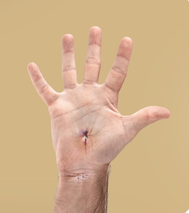
Endoscopic Carpal Tunnel Release is a minimally invasive surgical technique used to treat Carpal Tunnel Syndrome by relieving pressure on the median nerve.
This procedure consists of accessing the interior of the wrist with 1-2 small tubular devices, using smaller cuts than the traditional open surgery approach. A 2-3 cm cut is made on the wrist, and the endoscope (a tube with a camera attached) is positioned under the carpal ligament.
The cutting tools are guided through a second cut on the palm of the hand also measuring between 2-3 cm. The endoscope lets the doctor see structures in the wrist, most importantly the transverse carpal ligament, without having to create a large incision. These endoscopic methods have the advantage of sparing the skin on the hand and the tissues directly underneath, such as fatty tissues and the palmar fascia (a thick protective tissue). The transverse carpal ligament is cut, releasing the pressure on the median nerve. The cuts are then closed with stitches.
Usually, there is a more rapid and complete return to work and usual activities, at least 1-2 weeks sooner than when compared to open surgery methods.
Disclaimer: This video contains graphic content involving a surgical procedure.
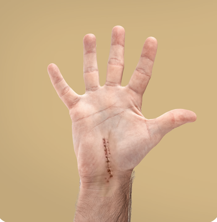
Mini-Open Carpal Tunnel Release is a traditional surgical method that requires an incision over the carpal tunnel area to directly access and cut the transverse carpal ligament.
A large 6-7cm cut is made on the palm of the hand and tissues below the skin are cut and moved aside so the doctor can see the transverse carpal ligament.
Once the ligament is cut, the pressure in the nerve is released. The tissues scar up deeply and superficially. This method allows the doctor to identify the structures through direct vision, avoiding injury to important structures. To reach the transverse carpal tissue (carpal tunnel tissue) which lies deeper, the doctor must cut through fatty tissues and a thick protective tissue called the palmar fascia. This can result in a thicker and more sensitive scar.
This approach leads to longer recovery & scarring compared to the other two options. The usual time for the tissue to heal is 6-8 weeks, with the scar typically taking longer.
Disclaimer: This video contains graphic content involving a surgical procedure.